Technological development and science are key at a sports level, especially in injuries. MRI turns out to be a fundamental tool for diagnosing muscular injuries and for the prevention and consequent development of the path that must be followed, especially in sports clinic cases.
Let's discuss what is new about MRI that makes it more effective than other techniques focusing on its scope and potentialities!
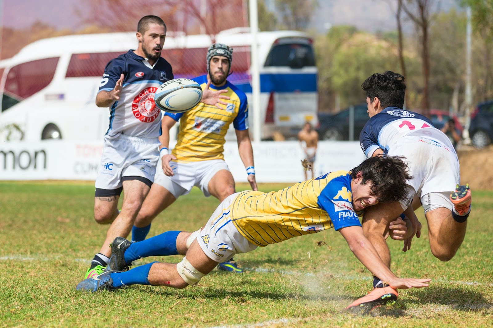
What Application does the 3T MRI have at a Sports Level?
Muscle injuries are prevalent in sports. In addition to causing long periods of inactivity, they can also be severe for the athlete, mainly if they occur during a critical moment of competition.
Studies show that muscle tears are the most frequent sports injury in Olympics, after ankle sprains, with 30% of all injuries caused during sports activity. The implications for the elite athlete, coach and team range from physical and psychological to financial issues.
Both plain radiology (Rx) and computed tomography (CT) have a limited role in detecting muscle injuries, especially in the acute stage. They can be useful methods for locating calcifications in cases of chronicity, sequelae of muscle injury with fibrous scarring, or when myositis ossificans is suspected of a muscle tear complication.
The scope of ultrasound may also be limited depending on the case. There are several deeper muscle damages, which are more infrequent but go unnoticed compared to ultrasound. MRI is of fundamental importance due to its scope and potential in this context.
3T MRI is the test of choice in this regard. It improves the diagnosis, treatment, and follow-up of neurological diseases because it allows us to see images and record the brain's neuronal activity.
Magnetic resonance imaging (MRI) stands out for its high-resolution image and the contrast of soft tissue (fat, muscles, ligaments, etc.), making it the ideal technology to diagnose sports injuries. It makes the diagnosis of injuries easier that are not easy to perceive through other systems, such as ultrasound, CT or X-rays. Muscle and ligament injuries, contusions and small bone fractures are the types of injuries in which getting an MRI scan can be most helpful.
Due to its high anatomical resolution and multiplanar capacity, MRI is the method of choice to detect deep or atypical muscle lesions. It also has a high sensitivity to provide high-contrast images in soft tissues and evaluate deep planes and a high specificity for the characterization of a muscle tear with infrequent imaging characteristics when its differential diagnosis with a primary infiltrative process is considered.
Accurate Information
MRI is considered the gold standard for diagnosing rare and deeper sports injuries. This is because it can describe the complexity of the conditions, regardless of their temporal evolution. MRI is also chosen due to its maximum sensitivity for detecting minimal lesions.
In sports medicine, the most outstanding achievements of MRI can be measured by its ability to provide concrete and measurable data. With them, doctors can estimate, for example, the recovery periods of athletes who present muscular injuries in the lower extremities, as well as the potential risk of a new injury.
MRI evaluation capacity is complemented by its non-invasive format, better temporal resolution, repeatability and higher precision. In addition, it has an excellent ability to provide high-contrast images in soft tissues and to evaluate deep planes. It also offers high specificity for coding a muscle tear with unusual imaging features.
Uncommon lesions detected with MRI
Studies show that MRI can detect infrequent lesions of:
· The upper limb with compromised muscles, such as the deltoid and coracobrachialis, in patients associated with boxing, rugby, weightlifters and throwers.
· The thoracic and abdominal walls detected in the pectoralis major, internal oblique and Quadratus Lumborum diagnosed in rugby athletes, weightlifters and runners
· The lower limb in Vastus Lateralis, Soleus, Flexor Hallucis, present in amateur soccer players
· The pelvic girdle and lumbar region with compromised muscles, such as gluteus medius, gluteus minimus, internal and external obturator and quadratus femoris, in aerobic athletes and marathon runners
MRI: Efficient and Precise
The evaluation of sports injuries in such a way would not have been possible without MRI. The anatomical resolution and multiplanar capacity provide precision in diagnosing the affected muscle group and provide tools to plan treatment and physical rehabilitation.
Likewise, the possibility offered by magnetic resonance imaging to differentiate tissues according to the weighted sequence and rule out other pathologies with a similar clinical presentation is beneficial.
What is the Mechanism of Resonance?
Magnetic resonance is a technology that does not use X - rays. Therefore, it is an innocuous technique since it avoids the negative effects of ionizing radiation. The images are obtained thanks to a magnetic field and a radiofrequency pulse.
It is done by activating the protons present in our body (generally the hydrogen proton present in the water in our body.) The relaxation of these protons emits a signal captured by an antenna near the patient. It is then digitized and transformed into an image that can be seen on a monitor or screen.
3T MRI technology applied to sports medicine has reached high standards, not only for the athlete's recovery but also for optimizing the care and treatment of their body on the way to better performance.
Loading comments...